Microglia and Macrophages in Brain Tumour biology
RESEARCH FOCUS
Numerous works, including ours, have emphasized the potential of activated tumour-associated microglia/macrophages (TAMs) as targets for anti-glioblastoma therapies. Tumour cells, but also tumor-initiating cells, regulate these immune cells and trigger their tumour-supportive activities. Our objectives are to characterise the nature of the signals that underlie this regulation, with a special emphasis on macrophage immunometabolism and metabolic changes taking place during cancer progression (e.g. glutamate and sphingolipid metabolism). We test the possibility to translate our results into potential therapeutic strategies by using the three-dimensional spheroid culture system.
Keywords: microglia, macrophages, TAMs, glioblastoma cells, glioblastoma initiating cells, inflammatory response, sphingolipid metabolism, glutamate metabolism, immunometabolism, normoxia, hypoxia, “omics” and bioinformatics.
COLLABORATORS & ONGOING PROJECTS
-Prof. Miguel Andrade, University JGU-Mainz and Institute of Molecular Biology, Mainz. Investigation of glutamate signalling and inflammatory response using computational biology and data mining.
-Dr. Laura Bindila, Medical University JGU-Mainz, Mainz. Lipidomic analysis.
-Prof. Bertrand Joseph, Karolinska Institute, Stockholm, Sweden. Role of DNA methyltransferases in microglia-tumor cells interactions
-Dr. Ella Kim, Medical University JGU-Mainz, Mainz. TAMs and Glioma Initiating Cells interactions; Glioma Initiating Cells response to drug combination.
-Prof. Javier Mora Rodrigez, Faculty of Microbiology, Universidad de Costa Rica, Costa Rica. Joint research initiative on the role of IL-38 as a novel immune regulator in the tumor microenvironment of glioblastoma multiforme.
-Prof. Rodrigo Mora, Faculty of Microbiology, Universidad de Costa Rica, Costa Rica Role of complex networks of microRNA-transcription factors in the regulation of the response of tumor-associated microglia/macrophages to glioblastoma-derived glutamate
-Dr. Gernot Poschet, Metabolomics Core Facility, Heidelberg. Metabolomics of TAMs.
-Prof. Wolfram Ruf, Medical University JGU-Mainz, Mainz. Macrophage metabolism
-Prof. Michel Salzet, Proteomics, Inflammatory Response and Mass Spectrometry Laboratory (PRISM), University of Lille 1, Villeneuve d’Ascq, France. Proteomics and Maldi-Imaging of glioblastoma spheroids
-PD Dr. Nicolai Savaskan, Neurochirurgische Klinik Universitätsklinikum Erlangen, Erlangen. Role of glutamate in microglia-glioma cells interactions
CURRENT ACTIVITIES
-Our projects-
Therapeutic manipulation of microglia/macrophages – targeting their immunometabolism
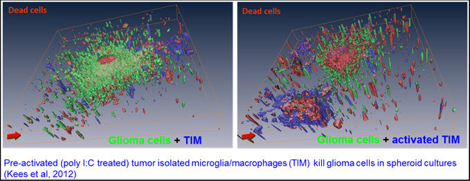
Microglia/macrophage-targeted therapy represent an interesting alternative or complementary therapy to classical therapies that target the glioblastoma (GB) cells. Strategies could aim at eliminating these tumor-supporting cells or at re-activating their cytotoxic potential. We have shown that stimulation of tumor-associated microglia/macrophages (TAMs) through their Toll-like receptors triggered a potent, tumor-specific cytotoxic answer that led to elimination of TNF- and TRAIL-resistant glioma cells and of temozolomide-resistant glioma cells. Experiments performed with TAMs and tumor cells co-cultured in spheroids showed that TAMs became fully refractory to this stimulation as a consequence of their growth in presence of glioma cells (Mora et al., 2009; 2010; Kees et al., 2012; Cisneros et al., 2016; Oancea Castillo et al., unpublished).
Based on this work and changes we observed in the glutamatergic signaling of TAMs in a glioblastoma environment (Choi et al., 2015), we started to characterize the metabolome of co-cultures of human monocyte-derived macrophages (MDMs) and glioma cells, its correlation with the inflammatory status of macrophages and its functional relevance for glioma growth and invasion. For that purpose, we undertook a multi-omics approach by combining analysis of extracellular metabolites, gene expression of a set of selected genes and proteomics of co-cultured MDMs. Two candidates (an enzyme and a glutamate transporter) were identified as potential targets and are currently under investigation through loss/gain of function analyses (Geiß et al.; coll. with N. Savaskan, G. Poschet; unpublished).
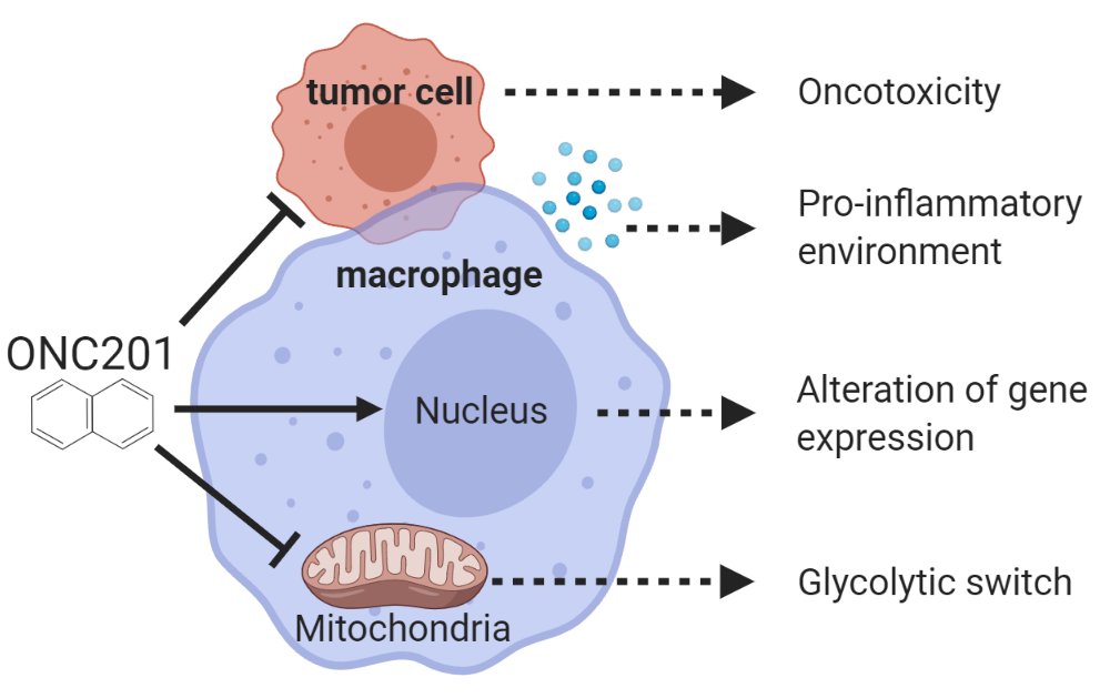
ONC201/TIC10 is an anti-cancer molecule which has emerged as a very valuable drug in cancer therapy and has been included in clinical trials for glioblastoma. It antagonizes the dopamine receptor D2 and affects mitochondria integrity in tumor cells. Its effects in TAMs are still ill-defined although ONC201 properties speak for possible effect on macrophage metabolic status. We examined whether ONC201 induces a metabolic and pro-inflammatory switch in primary human monocyte-derived macrophages that reactivates their anti-tumor activities, thus enhancing the onco-toxicity of ONC201. Macrophages responded to ONC201 with a severe loss of mitochondria integrity, a switch to glycolytic ATP production, alterations in glutamate transport and a shift towards a pro-inflammatory profile. Treatment of macrophages-glioblastoma cells co-cultures with ONC201 induced similar alterations in glutamatergic and inflammatory gene expression profiles of macrophages. It induced as well metabolic changes and a pro-inflammatory switch of the co-culture milieu. However, these changes did not translate into increased onco-toxicity (Geiß et al., 2021).
This study provides first evidence that ONC201 affects macrophage immunometabolism and leads to a pro-inflammatory tumor environment. The lack of increased cytotoxicity prompted us to investigate the effects of combining ONC201 with other molecules targeting tumor metabolism, an issue which is under current investigation in our lab (Geiß et al.; unpublished).
Evaluation of drug combination in vitro: efficient killing of glioblastoma cells and glioma initiating cells by a sphingosine kinase inhibitor combined with temozolomide under hypoxia
The standard treatment for newly diagnosed cases of glioblastoma is surgical resection followed by temozolomide (TMZ) chemotherapy combined with radiotherapy. However, patients commonly develop chemotherapy resistance, leading to a poor treatment response and tumor relapse. Sphingosine kinases (SKs), SK1 and SK2, are enzymes controlling the balance between two bioactive sphingolipids: the pro-survival sphingosine-1-phosphate (S1P) and the pro-apoptotic Ceramide. Elevated levels of S1P were found in GBM tissues and high SK1 expression correlated with a shorter survival time of GBM patients. Therefore, blocking SKs through pharmacological inhibitors has revealed to be a promising anti-cancer strategy. We demonstrated that combination of TMZ with a SK inhibitor does enhance cell death of GB cells (Noack et al, 2014).
We are now evaluating the efficiency of this combination under conditions that better mimic the tumor microenvironment by exposing GB cells to hypoxia. We determined a low-dose synergistic combination of TMZ and of the sphingosine kinase inhibitor SKI-II that specifically killed GB cells, but not normal human astrocytes, under hypoxia. We are currently characterizing the cell death mechanisms induced by this combination as well as its interplay with sphingolipid metabolism under normoxia and hypoxia. Effects on the invasion capacity of the GB cells is analysed as well. (N. Sousa et al.; coll. with L. Bindila; unpublished).
Glioblastoma recurrence is the biggest threat for patients. Thus, therapy should not only target tumor cells and TAMs but also glioblastoma stem cells (GSCs). This subset of cells possesses some characteristics of stem cells and is endowed with the capacity to initiate and propagate heterogeneous cancer cell populations. Would the TMZ/SKI-II treatment be able to prevent the dedifferentiation process supported by primary chemotherapy, it would most likely succeed in reducing tumor recurrence. This is what we currently investigate, analysing the response of GSC cell lines for their capacity to self-renew and differentiate under the treatment (N. Sousa et al.; coll. with E. Kim; unpublished).
-Our models of investigation-
Studying TAMs in appropriate in vitro experimental conditions
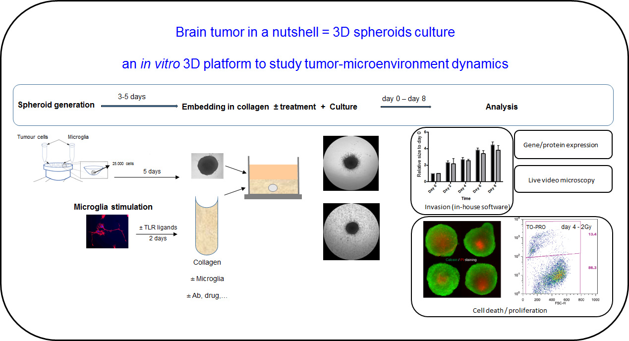
All our studies are in vitro studies, based on co-cultures of TAMs and GB cells. As in most laboratories worldwide, these in vitro studies are performed with cells cultured as monolayers in the atmospheric, i.e. 20- 21% oxygen normoxic conditions. Although these systems offer a fast read-out for cell response to treatment, they have various limitations such as: the high level of oxygen and their 2-D structure, which obviously are not reflecting the in vivo conditions of growth (low oxygen level) and morphology (3-D structure) of the tumor mass.
To integrate these elements in our current studies, we have started to perform experiments at low oxygen when we analyse monolayers of cells. Besides, we have implemented the use of cultures of spheroids. The use in cancer research of these complex three-dimensional (3-D) cell culture systems that mimic the in vivo human tissue dynamics has increased in the recent years. At diameters larger than 200 µm, tumor cell spheroids establish several chemical gradients (such as oxygen gradients) and thus different phenotypical and environmental layers (such as an inner hypoxic region), resembling those observed in genuine tumors. We have established a reliable, robust co-culture system that allows analysis of glioma cells-TAMs interactions and we developed a new software that facilitates an experimenter-independent evaluation of invasion (Cisneros et al., 2016). The co-culture system consists of spheroids (generated with tumor cells alone or mixed with microglia, TAMs or macrophages) implanted in a collagen matrix containing microglia (or macrophages, or TAMs). Cells can be treated before implantation into the collagen to modify their phenotype and function. Drugs can be applied at the beginning of the co-culture. Invasion, cell death, cell proliferation, gene expression and signalling can be analysed in this model.
Related publications
Mora, R., et al. (2009) TNF-α- and TRAIL-resistant glioma cells undergo autophagy-dependent cell death induced by activated microglia. Glia, 57, 561-581.
Mora, R., et al. (2010) Sphingolipid rheostat alterations related to transformation can be exploited for specific induction of lysosomal cell death in murine and human glioma. Glia, 58, 1364-1383
Kees, T., et al. (2012) Microglia isolated from glioma patients gain anti-tumor activities upon poly (I:C) stimulation. Neuro-Oncology, 14, 64-78. Epub 2011 Oct 20
Noack, J., et al. (2014) A sphingosine kinase inhibitor combined with temozolomide induces glioblastoma cell death through accumulation of dihydrosphingosine and dihydroceramide, endoplasmic reticulum stress and autophagy. Cell Death and Disease, 5, e1425; doi:10.1038/cddis.2014.384
Choi, J., et al. (2015) Glioblastoma cells induce differential glutamatergic gene expressions in human tumor-associated microglia/macrophages and monocyte-derived macrophages. Cancer Biology & Therapy, doi: 10.1080/15384047.2015.1056406.
Cisneros, L., et al. (2016) Evaluation of Consistency in Spheroid Invasion Assays. Sci. Rep., 6, 28375; doi: 10.1038/srep28375.
Cisneros, L., et al. (2016) A Novel Computer-Assisted Approach to evaluate Multicellular Tumor Spheroid Invasion Assay. Sci. Rep., 6, 35099. doi: 10.1038/srep35099.
Geiß, C., et al. (2019) Assessing the reliability of gene expression measurements in very-low-numbers of human monocyte-derived macrophages. Sci. Rep. 9, 17908. doi.org/10.1038/s41598-019-54500-8 1
Geiß, C., et al. (2021) Metabolic and inflammatory reprogramming of macrophages by ONC201 translates in a pro-inflammatory environment even in presence of glioblastoma cells. Eur.J.Immunol., DOI: 10.1002/eji.202048957